Geoffrey D. Rubin, M.D., Stanford Professor of Radiology and Diagnostic Radiology, discusses the various radiology technologies, their uses, and the positive changes they are making in medical diagnosis.
He begins with visual imagery of a woman who tripped and fell onto one of her knitting needles. We see a CT scan that displays her chest from top to bottom in a rotating set of images. We also see the same information rendered into a 3D graphic. By looking at these images one can see that the needle has penetrated her heart (right ventricle) and become aware of the true extent and location of her injuries. Additionally, the sharp eye of the radiologist, while diagnosing the chest injury, also noticed that her breast cancer had returned. With the assistance of these imaging techniques, the clinical staff was able to treat both of her medical ailments and she is doing well today.
Imaging=seeing into the living body. It's an advancement over prior techniques which relied on physical examination, listening, surgical examination and microscopic examination of tissue. These techniques are nice, but they fail to fully capture the body in action.
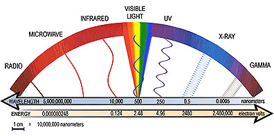
Imaging is done with energy. There are 3 ways:
1. Transmission. The passing of energy through the body with a visual examination of what comes out on the other side. This is the basis of radiography (X Rays) and computed tomography (CT).
2. Reflection. Energy is transferred into the body and then a measure is taken of the energy reflecting back out. This is the basis for sonography (Ultrasound).
3. Emission. The energy that is coming out from the body itself is measured. This is the basis for magnetic resonance imaging (MRI). It is also the basis of nuclear imaging - single photon emission tomography (SPECT) and positron emission tomography (PET).
X-Rays
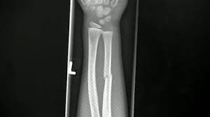
Here's how they work. An X-Ray beam is shined on the subject. The shining photons penetrate into the body and encounter the body's components mainly (carbon, nitrogen, hydrogen and oxygen, along with calcium and iron). The shining intensity is reduced as it encounters the body's parts, some of which are denser than others. The beam is then captured on the other side by either a fluorescing screen or a digital detector that converts the X-Ray into electrical signals. Irregularities can be seen as a result of uneven elements in the body, such as a broken bone, which will absorb less of the beam than surrounding items did.
Imaging=seeing into the living body. It's an advancement over prior techniques which relied on physical examination, listening, surgical examination and microscopic examination of tissue. These techniques are nice, but they fail to fully capture the body in action.
Imaging is done with energy. There are 3 ways:
1. Transmission. The passing of energy through the body with a visual examination of what comes out on the other side. This is the basis of radiography (X Rays) and computed tomography (CT).
2. Reflection. Energy is transferred into the body and then a measure is taken of the energy reflecting back out. This is the basis for sonography (Ultrasound).
3. Emission. The energy that is coming out from the body itself is measured. This is the basis for magnetic resonance imaging (MRI). It is also the basis of nuclear imaging - single photon emission tomography (SPECT) and positron emission tomography (PET).
X-Rays
Here's how they work. An X-Ray beam is shined on the subject. The shining photons penetrate into the body and encounter the body's components mainly (carbon, nitrogen, hydrogen and oxygen, along with calcium and iron). The shining intensity is reduced as it encounters the body's parts, some of which are denser than others. The beam is then captured on the other side by either a fluorescing screen or a digital detector that converts the X-Ray into electrical signals. Irregularities can be seen as a result of uneven elements in the body, such as a broken bone, which will absorb less of the beam than surrounding items did.
X-Rays were discovered by William Roentgen in 1895. X-Rays are electromagnetic radiation.
There are 5 fundamental tissue densities to examine in the image.
1. Gas. Molecules scattered. Not dense. Dark on X-Ray image.
2. Fat.
3. Water.
4. Mineral. Almost always calcium. Denser. Light on X-Ray image.
5. Metal.
Mammography is for the examination of breast tissue for cancer detection. This is done through analysis of the relative mixture of fat, water and calcium to locate irregularities.
He shows an image of pneumonia and notes that the chest is ideal for X-Rays because of its high level of gas, so when an infection is present it is easier to see because other elements, such as fluid, replace the gas and provide a clear image on the X-Ray.
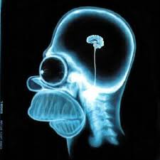
Due to the skull's density, projectional radiography is not able to provide insights into it. However, advanced technologies allow visualization of the brain's activities.
Computer technology is helpful but the computer can't really track and decipher the images the way that a trained radiologist can. For example, think of the difficulties faced by computers in recognizing human faces in motion.
CT - computerized tomography - captures a stack of cross sectional images that create a three dimensional image.
The images are taken from multiple angles and then combine into a three dimensional result. And this result can be created in seconds. CT scans are immensely helpful for diagnosing illness, including cancer detection and staging, trauma, abdominal injuries, stroke and vascular imaging.
Image processing is one of the biggest challenges for CT scans - how to review and interpret results quickly and accurately. This is an area of growth within computer processing of images, including better highlight rendering for visual inspection of specified areas.
Image processing is one of the biggest challenges for CT scans - how to review and interpret results quickly and accurately. This is an area of growth within computer processing of images, including better highlight rendering for visual inspection of specified areas.
